If the active selection is a single residue or ligand, the Details panel contains the following information:
Title
![]() |
The component name and 3-letter code. |
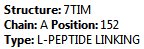
Summary
![]() |
- Structure – The name of the parent structure.
- Chain – The name of the parent chain.
- Position – The sequence position.
- Type – The component type.
|
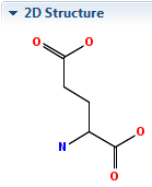
2D Structure
![]() |
The chemical structure for the component. |
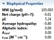
Biophysical Properties
![]() |
Displays the physical properties of the current selection, and may include:
- MW [g/mol] – The molecular weight of the selected component.
- Formal charge – The sum of formal charges in a molecule for a given electronic state. This is shown only for non-amino acid selections.
- Net charge – The pH-dependent sum of charges in a population of molecules. Protean 3D calculates the net charge for protein regions and amino acid residues, and assumes a pH of 7.
- pI – The isoelectric point (the pH at which the residue carries no net electrical charge), calculated from pKa tables from Lehninger et al. (2005). This is available only for amino acid selections.
- Average hydropathy – The Kyte-Doolittle hydropathy value, calculated using the method of Kyte, J. and Doolittle, R.F. (1982). This is shown only for amino acid selections.
- Aliphatic index – The relative volume occupied by aliphatic side chains (alanine, valine, isoleucine and leucine). See note below for reference. This is shown only for amino acid selections.
- A280 – The absorbance (optical density) for the residue, which is affected by presence of disulfide bonds. See note below for reference. This is shown only for amino acid selections.
- ε280; [M-¹cm-¹] – The extinction coefficient (amount of light a residue absorbs). Protean 3D assumes all Cys residues are reduced. See note below for reference. This is shown only for amino acid selections.
- Instability index – An estimate of the stability of the protein in a test tube. A protein whose instability index is smaller than 40 is predicted as stable, while a value above 40 predicts that the protein may be unstable. This value is calculated using the method of Guruprasad et al..
Note: For aliphatic index, absorbance and extinction coefficient, see Gasteiger et al.. In all three calculations, the sequence is treated as independent residues. There is no algorithmic dependence on length.
|
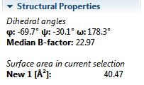
Structural Properties
![]() |
Displays angular and other structural properties of the current selection. A dihedral angle is the angle between two geometric planes defined by four atoms and is restricted by chemical and steric restraints.
Note: If the active document is a multi-model document, values are reported for the first model only.
- Dihedral angles φ (phi), ψ (psi), and ω (omega) are displayed only for proteins. φ and ψ define the structure of a protein’s backbone since ω is a rigid angle describing the peptide bond.
- Dihedral angles α (alpha) through ζ (zeta) and χ (chi) are displayed only for nucleotides. α through ζ are defined by the position of a residue’s backbone atoms while χ describes the orientation of the base.
- Median B-factor – The median temperature factor in a residue or ligand. For NMR files, this is typically 0.0.
- Occupancy – A list of alternate location codes and occupancies.
- Surface area in current selection is displayed only when a surface has been applied to all or part of a selection. The display shows the area, in square Angstroms, for the part of the surface that is contained in the selection.
|
Actions
![]() |
Click the relevant link to export data to a file, copy data to the clipboard, or open a browser window to the relevant page of the PDB or Ligand Expo databases. |